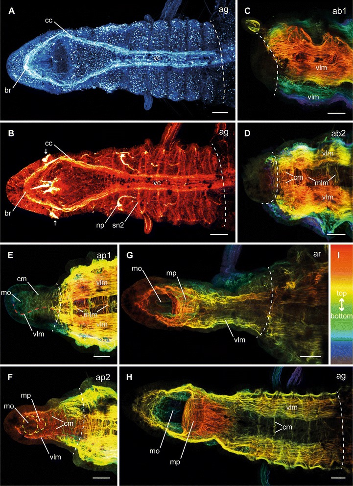
Fig. 4.

Different stages of anterior neuronal and muscular regeneration in Timarete cf. punctata. Anti-serotonin (cyan; A), anti-acetylated α-tublin (red; B), and anti-f-actine (depth coded, legend in I; C-H) staining, confocal maximum projections. Anterior is left, all views are ventral showing the anterior end, except C which is a latero-ventral view. The dotted white line indicates side of dissection. Regeneration process is staged according to Fig. 2, time after decapitation of correspondent specimen is given in brackets. a, b Growth stage (ag, 14 d). The nervous system almost reached its final shape. Aside from the size and the hardly definable segmental ganglia, the brain (br), the circumesophageal connectives (cc), as well as the ventral nerve cord (vc) with its segmental nerves (sn2) were present. c, d Early blastema stage (ab1, 48 h) and middle blastema stage (ab2, 72 h). The musculature still remained bluntly cutted without infiltration inside the blastema. e Early blastema patterning stage (ap1, 96 h). Thin circular (cm) and longitudinal muscle fibers (vlm) were visible inside the elongated blastema. The ventral longitudinal muscle fibers (vlm) were already organized in two strands, having an hourglass-like course (red dotted line). Further musculature started surrounding the mouth opening (mo). f Late blastema patterning stage (ap2, 5 d). The muscular elements became more prominent and the two strands of the ventral longitudinal musculature (vlm) diverged from each other to their final shape (dotted red line). The musculature of the mouth opening (mo) formed a pouch (mp) at its posterior edge. g Re-segmentation stage (ar, 9 d). The muscular pouch of the mouth opening (mp) continued its development and the ventral longitudinal musculature (vlm) now reached its final shape. h Growth stage (ag, 14 d). All muscular elements possessed an almost adult shape, now. Scale bars = 50 μm
