Fig. 5.

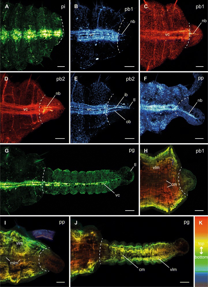
Different stages of posterior neuronal and muscular regeneration in Timarete cf. punctata. Anti-FMRFamide (green, A, G), anti-serotonin (cyan; B, E, F), and anti-acetylated α-tublin (red; C, D), as well as anti-f-actine (depth coded, legend in K; H-J) staining, confocal maximum projections. Anterior is left, all views are ventral showing the posterior end. The dotted white line indicates side of dissection. Regeneration process is staged according to Fig. 2, time after decapitation of correspondent specimen is given in brackets. a Invagination stage (pi, 48 h). b Early blastema stage (pb1, 60 h). Neurite bundles (nb), originated in the residual ventral nerve cord (vc) infiltrated the blastema. In this specimen, the outer neurite bundles grew faster than the inner ones. c Early blastema stage (pb1, 96 h). In this specimen, the inner neurite bundles grew faster than the outer ones. d, e Late blastema stage (pb2, 92 h). The neurite bundles have grown out. While in anti-acetylated α-tublin staining only a plexus of neurite bundels (nb) was visible, the anti-serotonin staining revealed inner (ib) and outer (ob) neurite bundles. The inner ones formed a loop (tl) in the most posterior position. f Blastema patterning stage (pp, 96 h). The neurite bundles (nb) represented a more compact structure, now. g Growth stage (pg, 12 d). The ventral nerve cord was well developed in the older regenerated segments, but faded the more posterior it run. h Early blastema stage (pb1, 96 h). Inside the blastema no muscular elements were detectable. i Blastema patterning stage (pp, 96 h). Also at this time point, there was no visible regeneration of musculature. j Growth stage (pg, 12 d). The older the segments were, the more developed the musculature was. In the younger segments, especially the circular musculature (cm) was rarely present. Scale bars = 50 μm
