Fig. 3.

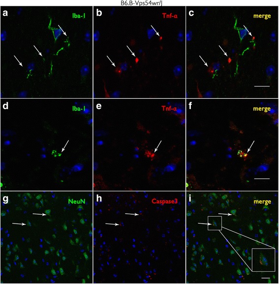
Tumor necrosis factor alpha (TNF-α) and Iba-1-labeled microglial cells and caspase 3-positive neuronal cells in motor cortex tissue of WR mice 40 d.p.n. a–f Iba-1-labeled microglial cells (green) also synthesize the cytokine TNF-α (red). f Microglial cells appear yellow due to the co-localization of Iba-1 (green) and TNF-α (red). Scale bar = 10 μm. g–i Caspase 3-positive (red) neurons labeled with NeuN (neuronal nuclei antibody) (green). Insert in i shows a caspase 3-positive neuron undergoing apoptosis. Scale bar = 20 μm
