
Fig. 6.
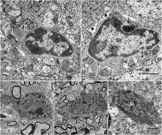
Ultrastructure of microglia and degenerating neurons in the motor cortex of B6.B-Vps54wr/J mice. a–e Electron micrographs from C57BL/6 J-Vps54 wr/wr mice 40 d.p.n. a A typical microglial cell with dense heterochromatin lining the nuclear membrane, a narrow rim of contrasting light cytoplasm, and slender microglial processes (arrows). b Microglial cell containing debris of unknown origin (arrow). These morphologic features are associated with phagocytotic cells. Asterisk: tagged astrocytic process. Scale bar = 5 μm. c–e Transmission electron micrograph of degenerating neurons. They show an irregular morphology with highly electron dense cytoplasm containing dense vacuoles, swollen mitochondria, and a dilated ER-Golgi network (arrow). The nucleus displayed highly condensed chromatin, clustered in peripheral bundles. Scale bar = 20 μm