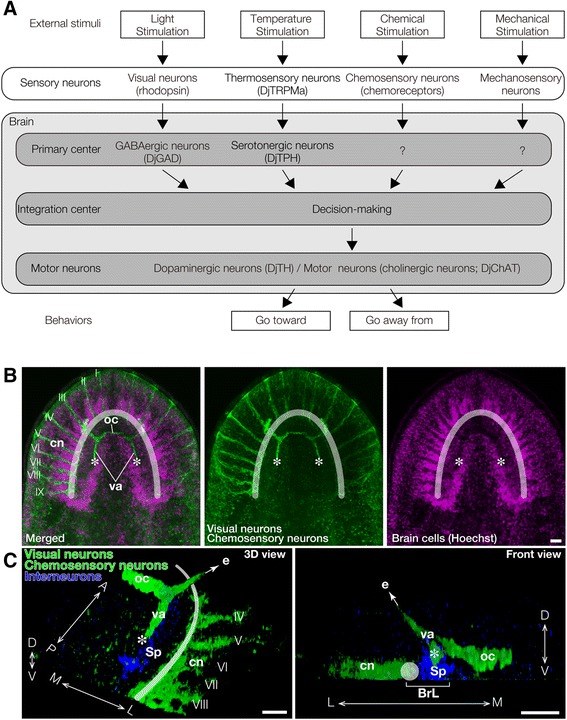
Figure 8.

Neural networks controlling planarian behaviors. (A) Planarians receive many kinds of signals from the outside, through independent types of sensory neurons such as rhodopsin-expressing visual neurons, DjTRPMa-expressing thermosensory neurons, and chemoreceptor-expressing chemosensory neurons, and these signals are processed by neurons in the brain, such as GABAergic neurons and serotonergic neurons. Thereafter, the various signals are integrated via certain neural networks in the brain to decide a planarian's behavioral strategy. Subsequently, the planarian behaves suitably in response to its conditions by controlling its motor neurons. (B, C) Different projections of visual neurons visualized by immunohistochemical staining with anti-arrestin antibody and chemosensory neurons visualized by staining with anti-DjGβ. (B) Axons of visual neurons form the optic chiasm and project to the medial region of the brain indicated by asterisks. Chemosensory neurons are located in nine lateral brain branches whose dendrites elongate toward the outer region of the head (I - IX) and whose axons project to the lateral region of the brain indicated by a diaphanous white arc. (C) 3D view (left panel) and front view (right panel) of visual neurons (green), chemosensory neurons (green), and interneurons in the spongy region of the brain (blue). Axons of visual neurons project to the dorso-medial region of the brain indicated by asterisks, whereas axons of chemosensory neurons project to the ventro-lateral region of brain indicated by a diaphanous white arc or circle. BrL, brain lobe; e, eye; cn, chemosensory neuron; oc, optic chiasm; Sp, spongy region; va, visual axons; A, anterior; P, posterior; M, medial; L, lateral; D, dorsal, V, ventral. Bars, 200 μm.
