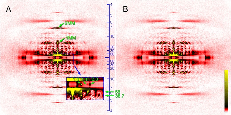
Fig. 3.

Diffraction patterns recorded from the flight muscle of Toxorhynchites towadensis. a relaxed; b rigor. The low-angle area of the diffraction pattern in (a) is magnified in the box on the lower right. In the box, the innermost equatorial reflections (1,0, 1,1 and 2,0) and the two lowest-angle layer-line reflections (d-spacing, 58 and 38.7 nm) are indicated by green arrows. Note that the 1st and 2nd myosin meridional reflections (1MM and 2MM) have single peaks right on the meridian. The scale on the right edge of panel a indicates d-spacing in nm
