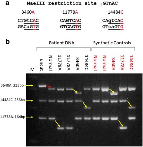
Fig. 2.

Multiplex PCR-RFLP. a: Diagram demonstrating the introduction of MaeIII restriction sites by the mutation (11778A) and the combination of primer alterations and mutation (3460A, 14484C). Mutations are shown in red with the primer alterations in lower case. b: 2.5 % ethidium bromide stained agarose gel showing the results of the PCR-RFLP on patient DNA (black labels) and synthesised DNA controls (red labels). DNA was PCR amplified in a multiplex reaction using 3460 F/R, 11778 F/R and 14484 F/R and restricted using 1 unit of MaeIII as described. M: size marker; Uncut: non-restricted PCR products; All other lanes: patient (Black) or synthesised DNA controls (Red) containing the indicated mutation PCR amplified and restricted with MaeIII. The red arrow between the uncut and normal lanes demonstrates the internal control of restriction. Yellow arrows demonstrate mutation detection
