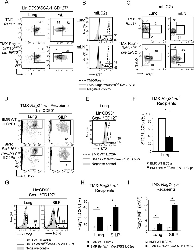
Figure 2.
The phenotypic alterations of Bcl11b−/− ILC2s are T cell independent and intrinsic. A) Flow cytometry analysis of the mILC2s and ILC3s in lung and mLNs of TMX-Rag1−/− Bcl11bF/F cre-ERT2 and -Rag1−/− mice. B) Flow cytometry analysis of surface ST2 in the mILC2s from lung and mLNs of TMX-Rag1−/−Bcl11bF/Fcre-ERT2 (black) and -Rag1−/− (dashed) mice. Gray shaded area represents negative control. C) Gata3 and Rorγt evaluated by flow cytometry in the mILC2s from lung and mLNs of the indicated groups of mice. Data (n=6) is representative of three independent experiments. D) Flow cytometry analysis of Lin−CD90+Sca-1+CD127+ ILCs in the lung and small intestine lamina propria (SILP) of TMX-Rag2−/−γc−/− mice reconstituted with bone marrow (BM) ILC2Ps (Lin−CD127+Sca-1hiST2+) from Bcl11bF/Fcre-ERT2 or wild type mice. Of note, donor mice were not treated with TMX, which was only administered to the recipient mice two weeks after transfer, as described in Figure S6A. E–F) Surface ST2 (E) and average frequency of ST2+ cells (F) in the Lin−CD90+Sca-1+CD127+ population from lung of TMX-Rag2−/−γc−/− mice reconstituted as in (D), with BM ILC2Ps from the indicated groups of mice. Gray shaded area represents negative control. G–I) Rorγt (G), average frequency of Rorγt+-cells (H), and average MFI of Rorγt (I) in the Lin−CD90+Sca-1+CD127+ population in the lung and SILP of the indicated groups of mice. Data (n=5), derived from two independent experiments, are presented as mean ± SEM. Significance was determined by Student's t test, * indicates p-value ≤ 0.05. See also Supplemental Figure S6.

