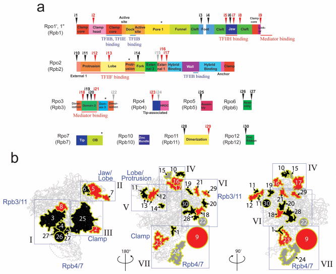
Figure 4. Structural differences between the Tko RNAP and yeast Pol II.
(a) Schematic diagrams of domains and domain-like regions of the Tko RNAP based on the Pol II nomenclature. Inverted triangles indicate positions of 30 insertions found in yeast Pol II compared with Tko RNAP (black, insertions in eukaryotic RNAPs; red, Pol II-specific insertions; gray, unique in yeast Pol II). The binding sites of TFIIB, TFIIE, TFIIF, TFIIH and Mediator are indicated. Asterisks indicate positions of 4 insertions found in the Tko RNAP subunits. (b) The structure of yeast Pol II and the three-dimensional representation of the 30 insertions in a. Insertions are depicted by plain-surfaces with yellow boundaries (colors same as in a). Circles show approximate locations of disordered insertions at N- or C-termini of subunits (e.g., Pol II CTD) and their diameters represent their approximate lengths. 7 groups of insertions (I to VII) are indicated in blue rectangles.

