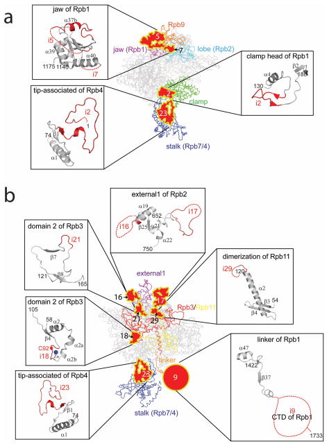
Figure 7. Structure and sequence comparisons of Tko RNAP and yeast Pol II around binding sites of the Pol II-specific GTFs/Mediator.
Structures of Pol II around the binding sites of TFIIF (a), TFIIH (b) and Mediator (c) and their counterparts in the Tko RNAP structure are shown. Insertions are colored and indicated in red and disordered regions are depicted as dashed lines. Amino acid sequence alignments around these regions are shown on their right (Tk, Tko; Ss, Sso; Sc, Sce; Hs, Homo sapiens). Amino acid residues cross-linked to TFIIF (a) and TFIIH (b) are indicated in red. Cys92 and Ala159 of Rpb3, which interact directly with Mediator, are indicated in red and the positions of i18 are indicated by a blue arrow on Tko RNAP and a blue circle on Pol II (c).

