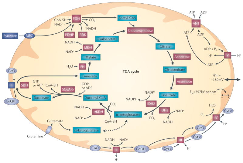
Figure 1. Mitochondrial bioenergetic function.
Pyruvate enters mitochondria via the mitochondrial pyruvate carrier (MPC), where it is decarboxylated and oxidized to acetyl-CoA by pyruvate dehydrogenase (PDH). Acetyl-CoA enters the tricarboxylic acid (TCA) cycle at citrate synthase. Subsequent steps in the cycle lead to the generation of reducing equivalents (NADH and NADPH) at dehydrogenase steps. Amino acids can also enter the TCA cycle by conversion to α-ketoglutarate, succinyl-CoA, fumarate, oxaloacetate or acetyl-CoA. For example, glutamine enters the TCA cycle after conversion to glutamate and subsequently to α-ketoglutarate. Reducing equivalents generated by the TCA cycle enter the electron transport chain and are eventually transferred to O2. Reducing equivalents generated at complex I (NADH–ubiquinone oxidoreductase) and complex II (succinate dehydrogenase (SDH)) are transferred to complex III by ubiquinol (CoQH2), a lipophilic quinone that carries a pair of electrons within the membrane. Electrons are transferred between complex III and complex IV by cytochrome c (cyt c), which carries a single electron coordinated to a haem group. Electrons that are transferred to complex IV (cyt c oxidase) are sequentially transferred to O2, generating H2O. Electron transfer steps at complexes I, III and IV are associated with proton translocation from the matrix to the intermembrane space, resulting in the generation of an electrochemical gradient across the inner membrane (ΔΨm). Complex V (ATP synthase) uses this gradient to catalyse the phosphorylation of ADP, yielding ATP. Exchange of ATP for ADP across the inner membrane is mediated by the adenine nucleotide transporter (ANT). Increases in ADP availability (as a result of cellular metabolic activity) tend to decrease ΔΨm slightly, which facilitates electron transfer at the steps involving proton extrusion. Hence, increases in cellular use of ATP cause an increase in mitochondrial respiration and ATP synthesis. By contrast, when ATP utilization falls, complex V activity decreases and the electron transport flux and oxygen consumption fall as a consequence of an increase in ΔΨm. Red highlighted proteins are known targets of reactive oxygen species (ROS). αKDH, α-ketoglutarate dehydrogenase; Em, electrical field within the membrane; FH, fumarate hydratase; IDH, isocitrate dehydrogenase; MDH, malate dehydrogenase; PDHP, pyruvate dehydrogenase phosphatase; PDK, pyruvate dehydrogenase kinase; Pi, inorganic phosphate; SCoA-S, succinyl-CoA synthase; SH, cysteine thiol.

