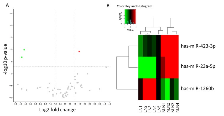
Figure 1.
Change of miRNAs in the LN metastasis group. (A) Volcano plot shows the fold change of miRNAs in the LN metastasis group compared to NLN group. The vertical lines correspond to 2.0-fold up and down respectively, and the horizontal line represents a P value of 0.05. The colored points in the plot represent the differentially expressed miRNAs with statistical significance. (B) Hierarchical clustering for differentially expressed miRNAs in the LN metastasis group versus NLN group using a volcano plot (fold change ≥ 0.5). Red indicates high relative expression and green indicates low relative expression.

