Figure 4.
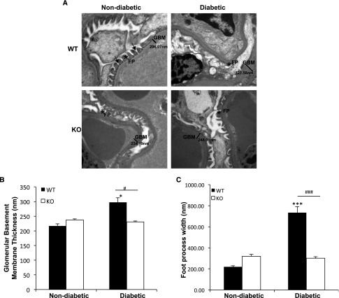
Diabetic TxNIP−/− mice do not develop podocyte foot process effacement and glomerular basement membrane thickening. Podocyte foot process effacement and GBM thickness in nondiabetic and diabetic TxNIP WT and KO mice. (A) Representative electron microscopic images (original magnification, approximately ×15,000) of glomeruli from control and diabetic TxNIP WT and KO mice. Arrows indicate slit diaphragm. FP, foot process. (B) Quantification of GBM thickness and (C) podocyte foot process effacement determined as described in the Concise Methods from nine different fields (n=3 mice/condition). Results are mean±SEM. *P<0.05 and ***P<0.001 diabetic WT mice versus nondiabetic WT; #P<0.05 and ###P<0.001 diabetic WT versus diabetic TxNIP KO mice.

