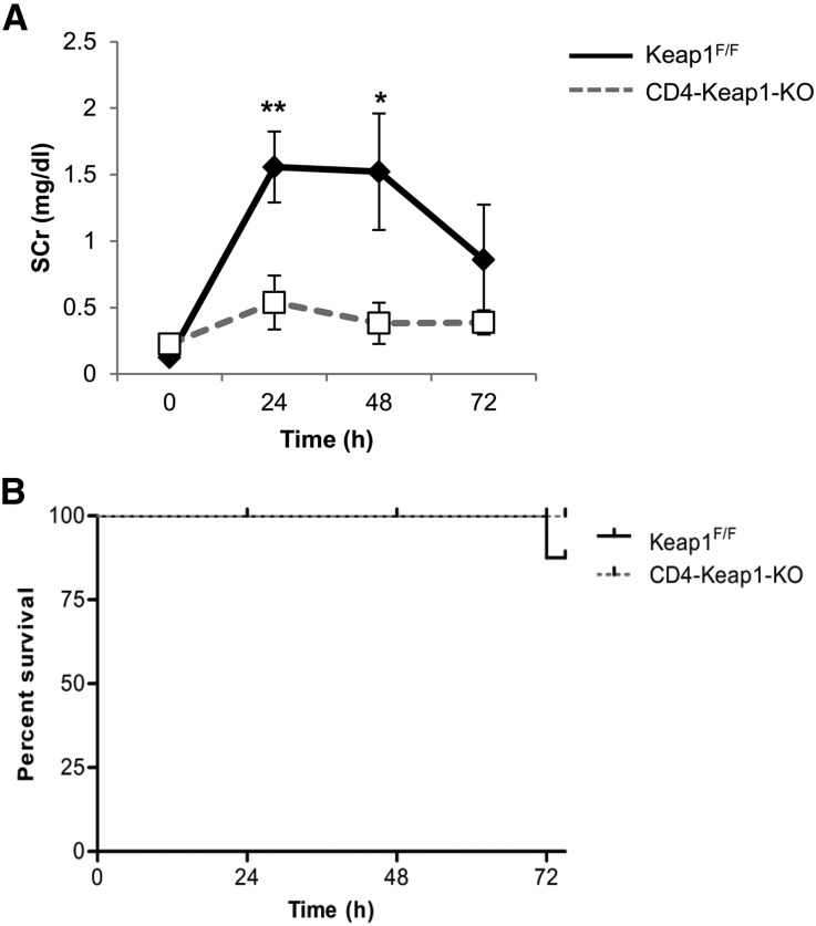
Figure 4.
Effect of T cell–specific Keap1 deletion on IR-induced AKI. (A) Deletion of Keap1 from T cells in CD4-Keap1-KO mice (n=7) improves kidney function after bilateral IR injury compared with Keap1F/F mice (n=9). (B) There is no mortality in CD4-Keap1-KO mice; however, 20% of mice died in the control group 72 hours after IR injury. (C) Representative images of hematoxylin and eosin–stained kidney sections showing significantly fewer necrotic tubules and greater normal renal cortex and medullary tissue in CD4-Keap1-KO mice compared with Keap1F/F mice 24 and 72 hours after IR injury. (D) Dot plot showing the percent score for necrotic tubules and normal cortex and medulla for CD4-Keap1-KO (n=8–10) and Keap1F/F (n=9–11) mice 24 and 72 hours after IR injury. (E) Proinflammatory cytokine IFN-γ is lower in whole kidney lysates of CD4-Keap1-KO mice compared with Keap1F/F mice 72 hours after IR injury, whereas TNF-α, MCP-1, and IL-10 are not significantly different between the groups. Graphs represent the mean±SEM. *P≤0.05; **P≤0.01. MCP-1, monocyte chemoattractant protein-1. Original magnification, ×200 in C.


