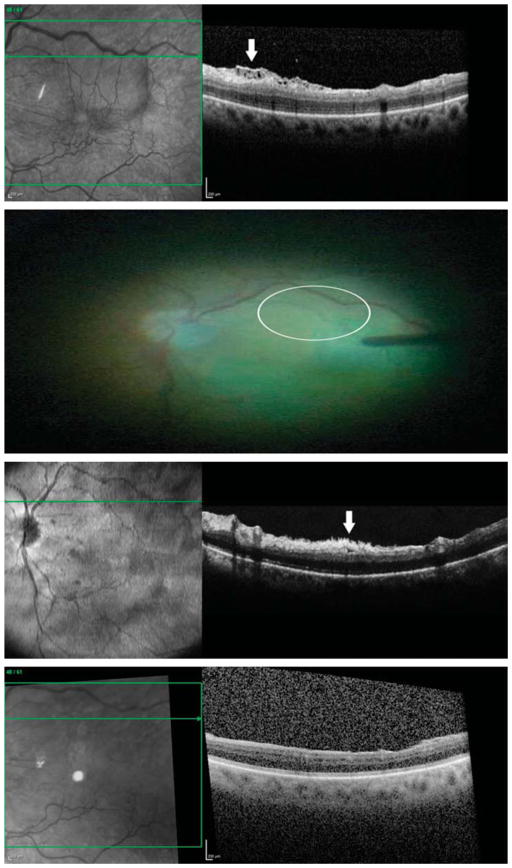
Fig. 3.
Optical coherence tomography images from a 61-year-old white man with an ERM accompanied by connecting strands. Preoperatively, a horizontal sectional image showed a moderately reflective, connecting lesion under a highly reflective, smooth ERM (arrow). The connecting strands had visible connections from the inner retinal surface to the ERM (first row). Before ILM peeling but after ERM removal, the ILM was already absent (it appeared to be removed along with the ERM removal in this area) as demonstrated by the lack of indocyanine green staining (circle) in the region of the preoperative connecting strands (second row). Although the ERM and the ILM have been removed, an intraoperative horizontal sectional image showed a highly reflective lesion (arrow). The shape of this lesion appeared as tufts. The remained disconnected lesions were contiguous with the inner retinal surface and seemed to originate from the inner retinal layers (third row). Three months after surgery, the intraoperative remaining lesion disappeared completely, and there was no recurrent formation of ERM (fourth row).

