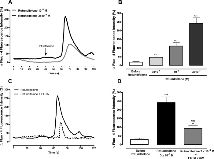
Fig 8. Rotundifolone-induced Ca2+ transient in myocytes depend of extracellular Ca2+.
(A) Representative recording of the Ca2+ signals induced by rotundifolone (10−3 and 3x10⁻³ M) in freshly dispersed vascular smooth muscle cells from rat superior mesenteric artery. (B) Summary data showing the amplitude of the Ca2+ signal induced after perfusion with different concentrations of rotundifolone (3x10⁻⁴, n = 9; 10−3, n = 5; 3x10⁻³ M, n = 11). (C) Representative recording of Ca2+ oscillations induced by rotundifolone (3x10⁻³ M) in myocytes placed in Ca2+-free medium fortified with 2 mM ethylene glycol tetraacetic acid (EGTA). (D) Summary data demonstrating the decreased amplitude of the Ca2+ signal induced by rotundifolone in Ca2+-free medium (n = 13). The Ca2+ oscillations were measured and represented as increases in fluorescence intensity relative to baseline [DF (%) = (F-F0/F0)*100]. The data were examined using unpaired Student's t tests (rotundifolone with or without EGTA) and one-way ANOVA followed by the Bonferroni post-test. (** p<0.01 and ***p < 0.001 vs control; ###p<0.001 vs Rotundifolone 3x10⁻³ M).

