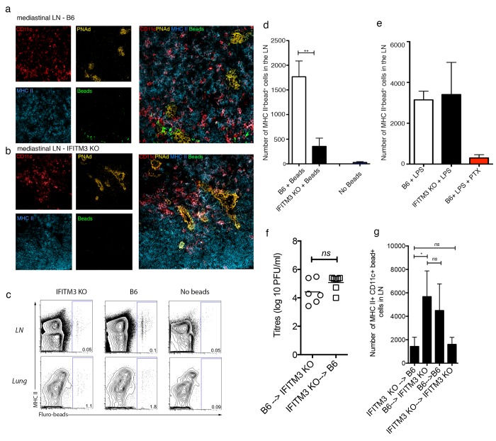
Fig 4. IFITM3 KO DCs are impaired in trafficking to the draining LN following influenza virus infection.
B6 or IFITM3 KO mice were intranasally instilled with fluorescent-beads and influenza virus. Lungs and lung-draining LN were harvested 48hrs later. Immunofluorescent staining of LN sections from (a) B6 and (b) IFITM3 KO mice at 48hrs post-infection. Staining for CD11c (red), MHC II (blue), PNad (yellow) and beads (green). x20 magnification, representative for 2 independent experiments (c) Representative flow cytometry profiles depicting beads within MHC II+ DCs in lung and LN. (d) The absolute number of bead+ MHC-II+ DCs in the draining LN (n = 5 mice per group, data pooled from 2 independent experiments, Student’s t-test **P <0.01) (e) B6 and IFITM3 KO mice were intranasally instilled with fluorescent-beads and LPS +/- pertussis toxin (PTX). The absolute number of bead+ MHCII+ DCs in the lung-draining LN was determined 48hrs later. Bars represent the mean + sem (n = 5, data pooled from 2 independent experiments) (f-g) B6—>IFITM3 KO, IFITM3 KO→B6, B6→B6 and IFITM3 KO→IFITM3 KO mice were intranasally instilled with fluorescent-beads and influenza virus. (f) Viral titres in the lung on day 2 post infection was measured by standard plaque forming unit assay (g) Lungs draining LN were harvested 48hrs later and the absolute number of bead+ MHC-II+ DCs was determined (n = 5–8 mice per group, data pooled from 3 independent experiments, Student’s t-test **P <0.01).

