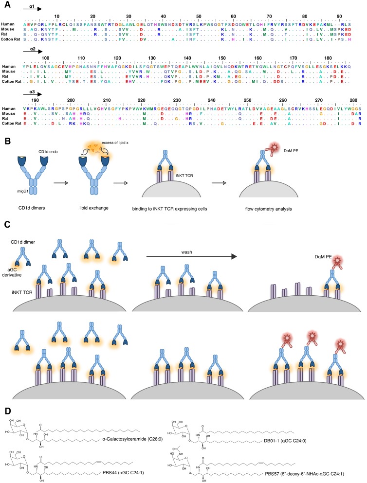
Fig 1. Experimental setup.
(A) α1 and α2 domain amino acids of CD1d molecules used in this study. Alignment was performed using BioEdit. Human: GenBank BC027926.1, Mouse: GenBank AK002582.1, Rat: GenBank AB029486.1, Cotton Rat: GenBank KM_267558. (B) In vitro loading analysis of CD1d dimers presented as schematic overview. (C) Scheme explaining the emergence of different saturating curves depending on loading efficacy of dimers. Upper panel: Inefficient loading leads to low proportion of functionally bivalent CD1d dimers, Lower panel: Efficient loading leads to high proportion of functional bivalent CD1d dimers. Drawing represents several step flow cytometry analysis of incubation/wash/detection. (D) Structural features of all glycolipids used in this study.

