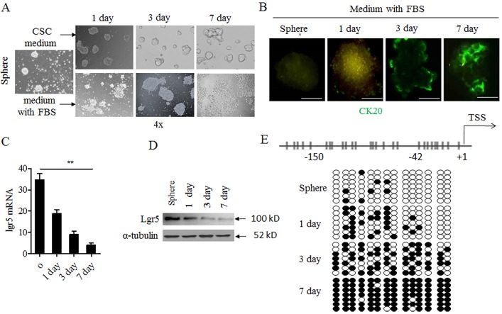
Fig 2. Lgr5 expression and methylation varied from CSC spheres to adherent cells.
A. CSC spheres from HCT116 cell lines grew with CSC medium without serum (upper panel); Or, CSC spheres changed to normal medium with 10% FBS (down panel). Observation time point: 1 day, 3 day. And 7 day and photographed under light microscope. B. Immunofluorescent staining of CK20 in CSC spheres; Green indicated ck20 staining. C. RNA was collected in different time point and was analyzed by qPCR in medium with 10% FBS group. D. The protein was collected in different time point in medium with 10% FBS group, lgr5 was detected with western blot.westernblot α-tubulin was used as internal control. E. BSP analysis of lgr5 promoter methylation density in CSC spheres and time point in in medium with 10% FBS group. Black circle, methylated CpG site; white circle, unmethylated CpG site.

