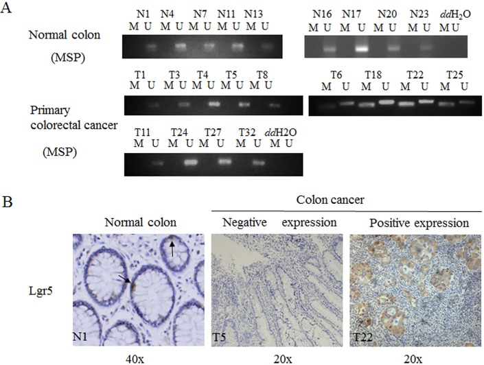
Fig 4. Lgr5 methylation was in normal colon tissues and primary colorectal cancers.
A. MSP was performed on normal colon tissues (A) or primary colorectal cancers. M, methylation signal; U, unmethylation signal. B. Lgr5 expression was in normal tissue and cancer tissues, dark arrows represented the lgr5 positive cell in by Immunohistochemical staining. N1 represented the one of the normal colon tissues; T5 and T22 respectively represented the colon cancer patients.

