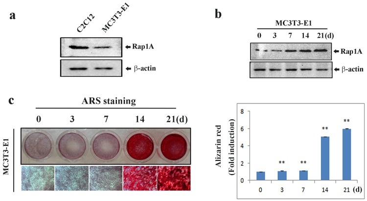
Fig 1. Expression of Rap1A during osteoblastic differentiation.
(a) Endogenous expression of Rap-1A in MC3T3-E1 and C2C12 cells by Western blotting. (b) The temporal change of Rap1A expression in MC3T3-E1 cells during osteoblastic differentiation by Western blotting. (c) Alizarin red staining for bone nodules in MC3T3-E1 cells. Original magnification was ×20 (left). Arizalin red-S staining activity was quantified by densitometry at 562 nm (right). Data represent means ± SD of triplicate samples. *P < 0.05, **P < 0.01 vs. the undifferentiated cells. β-actin was used as the internal control.

