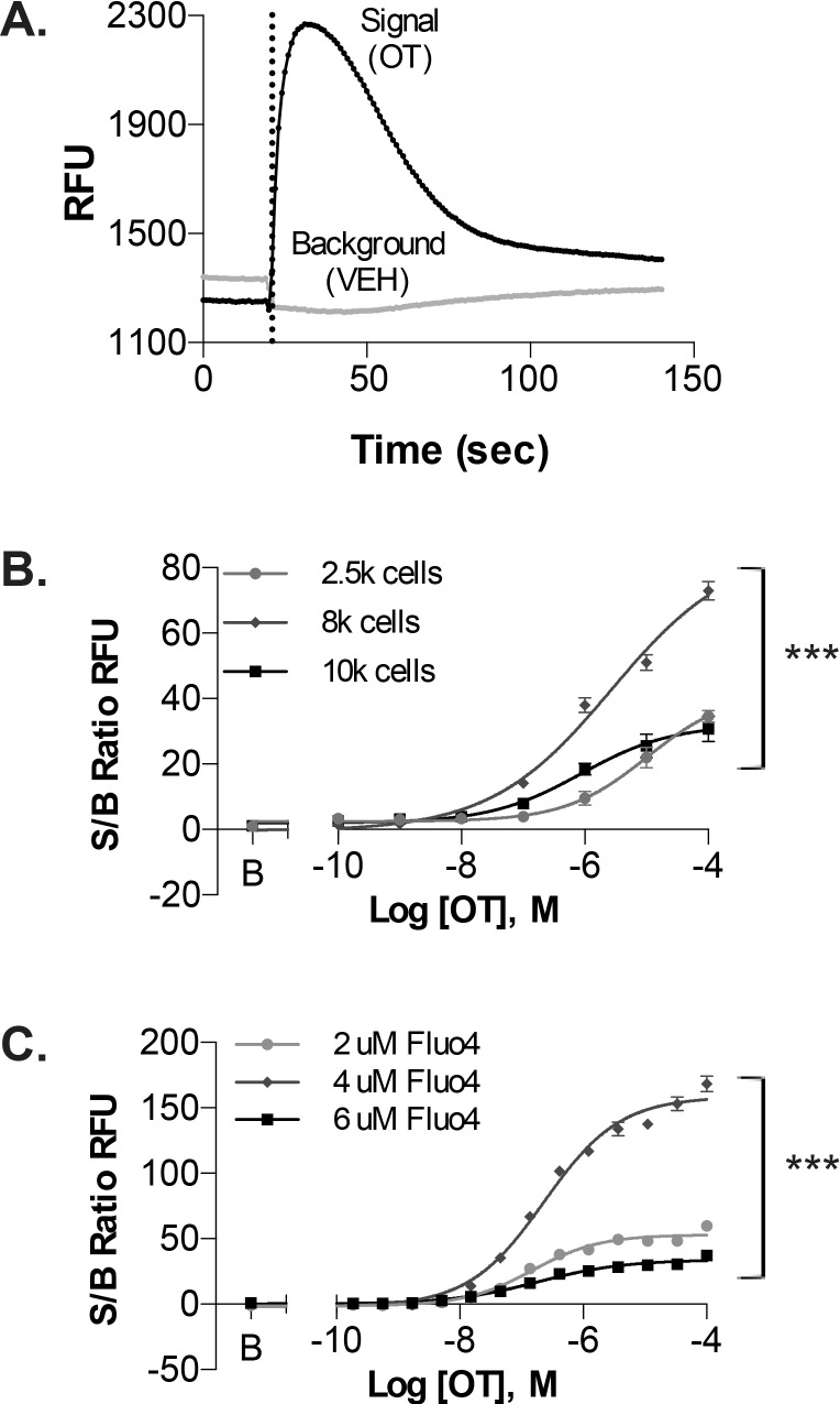
Fig 2. Ca2+-mobilization assay using uterine myometrial cells.
A. Representative recording of OT-induced Ca2+-mobilization from UT-myo cells loaded with Fluo-4AM. Ca2+-mobilization was monitored as an increase in Relative Fluorescent Units (RFUs). Dashed line indicates time of OT or vehicle (VEH) addition. Optimal assay conditions were determined by performing cell density gradient (B) and Fluo-4AM concentration response curves (C), from signal-to-background (S/B) ratios of Max-Min RFU obtained from OT and VEH wells, respectively. Non-linear regression was used to fit the data (Mean±SEM; n = 8 well replicates); significant (***p<0.0001) difference between each fit line.

