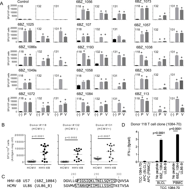
Fig 4. Validation of the IFN-γ response to selected HHV-6B peptides.
Peptide-expanded T cell cultures to each of the 14 peptides in Table 4 were generated for 3 donors, and tested for IFN-γ using ELISpot (SFU/106 cells). A. Responses of all expanded T cells to autologous PBMCs pulsed with the peptide (P) or HHV-6B (V). ELISpot negative controls are indicated by (-). Positive responses were assessed by DFR2x and are indicated by (+) on top of the bar. B. Summary of the responses of expanded T cells to autologous PBMCs pulsed with HCMV or HHV-6B for two HCMV seronegative (#118 and 132) and one seropositive (#131) donor (Table 1). P-values for HCMV vs HHV-6B (unpaired t-test) are shown. C. Alignment of homologous HHV-6B U57 and HCMV UL86 sequences in the region of known epitopes (boxes indicate the complete stimulating sequence). D. Response of a 6BZ_1084 CD4 T cell clone to autologous BLCLs or PBMCs pulsed with HHV-6B 6BZ_1084 or with HCMV UL86-B peptides, as measured by IFN-γ ELISA. P-values for no-peptide vs peptide (unpaired t-test) are shown.

