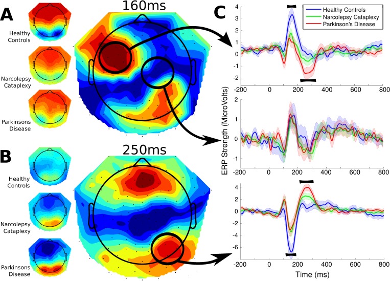
Fig 3. Feedback-locked group topographies and statistics at 160 and 250ms and Event-Related Potentials over right-posterior channels (the mean of 8 channels in the region).
Reward feedback could indicate a successful or unsuccessful trial. Significant differences were found around two principle time points indicated by the topographies. A, The first difference showed similarly low amplitudes for the two patient groups compared to the healthy controls and may correspond to the N170 (12 participants in each group). B, The second, later difference showed an ERP for patient groups which was virtually absent in healthy controls. Additionally, significantly larger amplitudes for this component were found for Parkinson’s patients compared to participants with Narcolepsy-Cataplexy. These differences may correspond to feedback-related negativity (FRN) C, Three individual time courses of the entire event-related potential. The top and bottom panels both show the positive and negative deflections of the two topographies, while the middle panel indicates a time course where no significant differences were found for reference.

