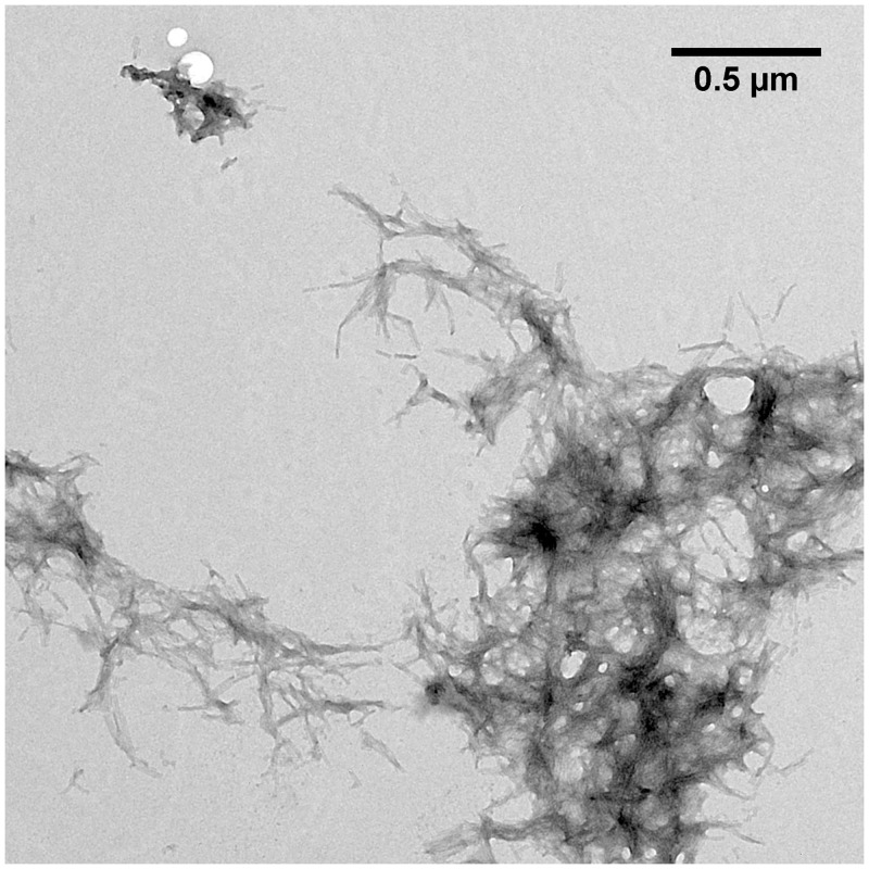
Fig 3. TEM image of pEAβ(3–40) in 50 mM potassium phosphate pH 2.8 containing 20% TFE.
Monomerized pEAβ(3–40) (25 μM) was incubated for fibrillation at 20°C for five days and grids were prepared by negative staining. pEAβ(3–40) incubated in aqueous TFE solution formed large twisted fibrils up to several hundred nm in size which accumulate into large aggregates ranging from 1–5 μm in diameter.

