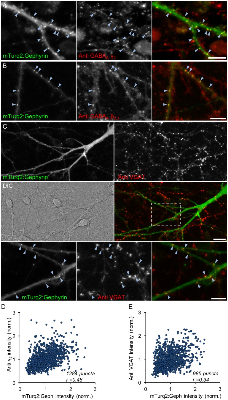
Fig 2. mTurq2:Geph colocalizes with GABAA receptors and presynaptic boutons of GABAergic neurons.
A,B) GABAA receptors were labeled in live hippocampal neurons using antibodies against extracellular epitopes of the GABAA receptor γ2 subunit (A) or GABAA receptor β2,3 subunits (B). Left panels: mTurq2:Geph; Middle panels: antibody labeling; Right panels: combined images. Note the good colocalization of mTurq2:Geph puncta with clusters of labeled receptors (arrowheads point to some examples). Bars, 10μm. C) Functional presynaptic boutons of GABAergic neurons were labeled in live neurons using anti vesicular GABA transporters (VGAT) antibodies. Top panels—mTurq2:Geph (left) and anti-VGAT (right). Middle panels—differential interference contrast (DIC) image of the same field (left) and the combined image (right). Bottom panels—enlarged images of region enclosed in rectangle in right middle panel. Note the good colocalization of mTurq2:Geph puncta with clusters of labeled VGAT (arrowheads). Bars, 20μm (middle), 10μm (bottom). D,E) Correlation between mTurq2:Geph fluorescence and GABAAγ2 labeling (D; 3 Experiments, 18 cells, 1284 synapses) and VGAT labeling (E; 4 Experiments, 17 cells, 985 synapses).

