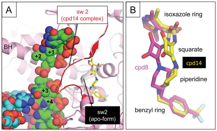
Figure 3.
(A) Comparison of the switch 2 conformations in the apo-form of RNAP (white) and bound with compound 14 (red). The switch 2 conformation when compound 14 is bound clashes with the template DNA of the RNAP-DNA complex. Structures of the apo-form RNAP, the RNAP – compound 14 complex and the RNAP elongation complex (template DNA, green; non-template DNA, cyan) were aligned by superimposing their β′ subunits. The DNA position of the template DNA and the RNAP bridge helix (BH) are labeled. Switch 2 is indicated in red with its disordered region shown as a dashed line. (B) Comparison of binding of compound 8 (magenta) and 14 (yellow) to the switch region of E. coli RNAP. Compounds are shown as stick models and their chemical groups are indicated.

