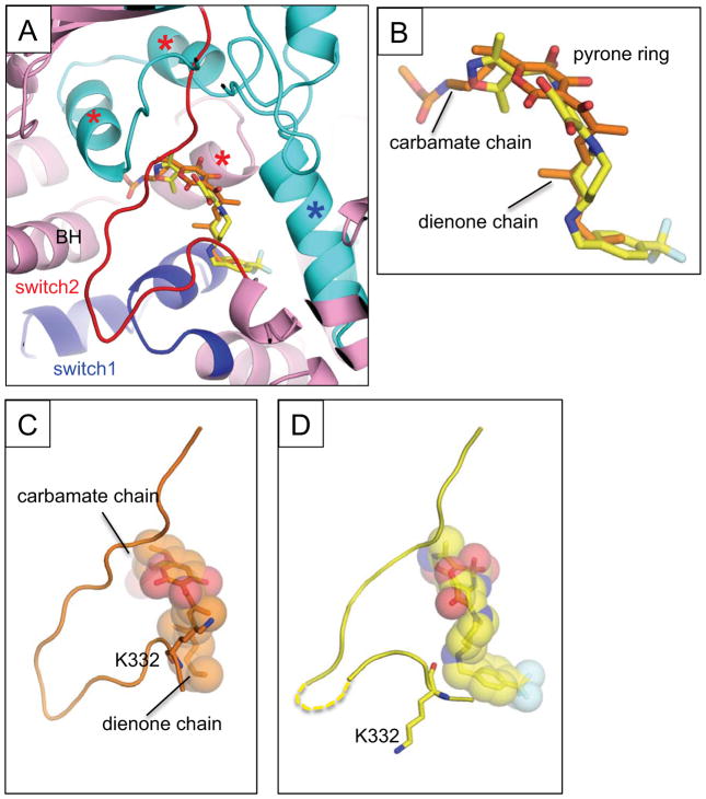
Figure 4.
(A) Comparison of the MyxB and squaramide bindings on the E. coli RNAP. Structure of the RNAP – MyxB complex was superposed with the RNAP – compound 14 complex. Only compound 14 is shown in the RNAP – MyxB complex structure. The β (cyan) and β′ (pink) subunit are depicted as a ribbon model, the switches 1 and 2 are indicated as blue and red, respectively. The MyxB (carbon atoms are shown in orange) and compound 14 (carbon atoms are shown in yellow) are shown as stick models. The three α-helix bundle contacting with the carbamate chain of Myx are indicated by red asterisks, the C-terminal α–helix of the β subunit interacting with the dienone chain is indicated by a blue asterisk. The RNAP bridge helix (BH) is labeled. (B) Comparison of the MyxB and compound 14 in the RNAP complexes. Orientation of this panel is the same as in (A). Chemical groups of the MyxB are labeled. (C–D) The switch 2 conformations of the E. coli RNAP – MyxB complex (C) and the E. coli RNAP –compound 14 complex (D).

