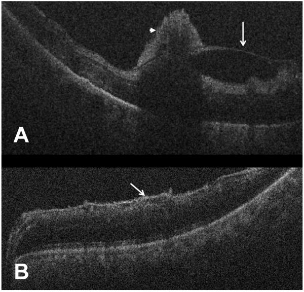
Figure 4.

Intraoperative optical coherence tomography demonstrating preretinal membranes following removal of vitreous hemorrhage. (A) B-scan shows macular edema, posterior hyaloidal traction (white arrow), and pre-retinal hemorrhages (arrowhead, Case 2). (B) B-scan reveals epiretinal membrane without significant retinal thickening (white arrow).
