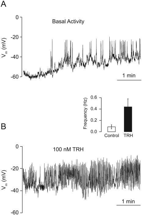
Fig. 1.
Characterization of spontaneous and TRH-stimulated electrical activity in TαT1 cells. (A) The electrical activity measured in current clamp mode in nystatin perforated patch showed a resting membrane potential of −55 ± 4 mV and spontaneous activity, characterized by small (< 10 mV; mean frequency 0.3 Hz) and big depolarizing events (> 10 mV; frequency 0.08 ± 0.03 Hz). (B) The TRH stimulation (100 nM) induced a depolarization of 10 mV and increased the firing frequency of the action potentials to 0.4 ± 0.1 Hz, with amplitudes of 35 ± 6 mV. (Inset) Frequency of spiking in unstimulated and TRH-stimulated cells.

