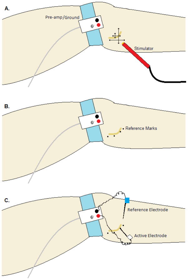
Figure 1. Typical layout and procedure for a peroneal (fibular) nerve recording.
The pre-amplifier and ground are attached to the skin on a flat surface either at the lateral knee joint (shown) or distal to the fibular head on the lateral shin. A) The systematic pattern of cutaneous electrical stimulation to locate and map the path of the nerve. B) The dots are placed on the skin following the path of the nerve and act as a reference for insertion of the recording electrode. 3) The reference electrode (blue) is placed beneath the skin and into the tissue within 2 cm of the expected recording site. The active electrode (white) is inserted through the skin and manipulated until a satisfactory nerve signal is acquired.

