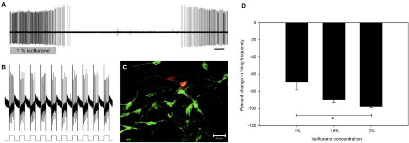
Figure 1. Medullary raphé 5-HT neurons are inhibited by isoflurane.
a) Representative extracellular recording of a 5-HT neuron that ceased firing during isoflurane treatment. The neuron returned to baseline firing rate upon washout. Under baseline conditions, firing frequency (1.12 Hz) and pattern was characteristic of serotonergic cells. Scale bar = 30 s. b) The same neuron was entrained to fire simultaneously with an applied stimulus and filled with biotinamide. c) Positive staining for TPH (green) and biotinamide (red) colocalize (yellow) to indicate the recorded neuron was serotonergic. Scale bar = 50 µm.

