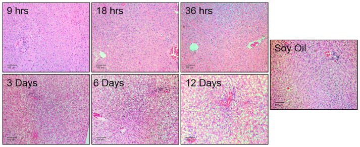
Figure 1. Progression of PCB126-mediated histological changes in the liver.
Male SD rats were treated with 5 umol/kg PCB126, intraperionteally at various time points resulting in exposures of 9 hr, 18 hr, 3 days. 6 days, and 12 days. Sections of the liver were stained with H&E stain and analyzed by a pathologist. These are representative images taken from each group. Scale bar equals 100 um.

