
Fig. 3.
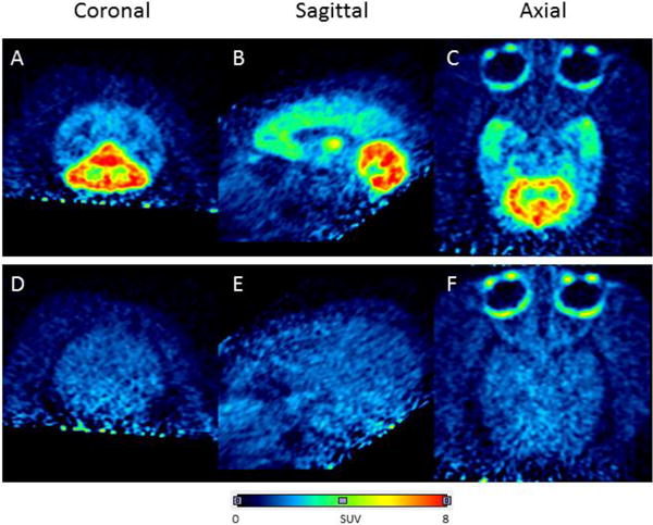
Brain PET images acquired as summed data from 0–90 min after intravenous injection of anesthetized rhesus monkey with [11C]FIMX (~ 200 MBq) at baseline (top row) and after preblock of mGluR1 with the selective antagonist JNJ16259685 (3 mg/kg, i.v.) (bottom row). Panels A and D are coronal, B and E, sagittal, and C and F transaxial images, respectively.