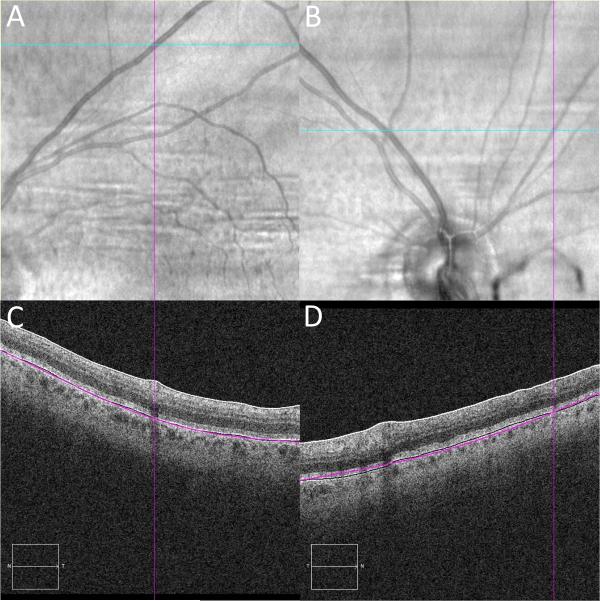
Figure 2.
IR and OCT B-scan alignment validation using retinal vessels
2A/B: IR image with the cyan line marking the section of OCT B-scan image and the purple line positioned over the center of the retinal vessel along that section. 2C/D: OCT B-scan image with vertical purple line marking the retinal vessel.

