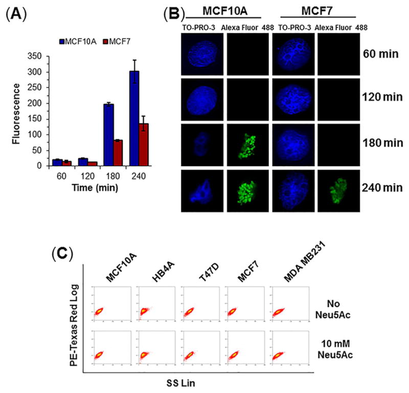
Figure 2. Optimization of nutrient depletion conditions.

(A) TUNEL assay for apoptosis. Cells were treated with 10 mM Neu5Ac for varying time points followed by treatment with BrdUTP and immuno-stained with anti-BrdU antibody-Alexa Fluor 488 conjugate and analyzed by flow cytometry. The level of fluorescence indicates the degree of DNA damage. (B) Cell imaging. Anti-BrdU Ab-Alexa Fluor 488 labeled cells were visualized under confocal microscope. Magnification: 100×. (C) Cytograms showing determination of live cells. Cells were treated with or without 10 mM Neu5Ac in nutrient deprived condition for 2 h and stained with 1 mg/mL propidium iodide (PI) on ice for 20 min and analyzed by flow cytometry in PE-Texas Red setting.
