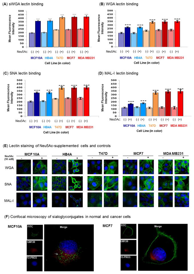
Figure 5. Flow cytometric analysis of lectin binding.
Cells were treated in the presence or absence of 10 mM Neu5Ac under nutrient deprivation for 2 h. The α2→3 and α2→6 sialylated glycans were stained with the fluorescein isothiocyanate-labeled lectins, sWGA (A), WGA (B), SNA (C), and MAL-I (D) followed by analysis on a flow cytometer. (E). Lectin staining followed by cell imaging. Cells were treated for 2 h in the presence or absence of Neu5Ac (10 mM) under nutrient deprivation and stained for 1 h with FITC-labeled lectins (WGA, SNA, and MAL-I) at concentration of 5 μg/mL (green fluorescence). Cells were further treated with 50 μg/ml ribonuclease A and the nuclei were counter-stained with TO-PRO-3 (blue fluorescence), Images are shown at 60x magnification. (F) Confocal imaging of MAL-I staining. Co-staining of Neu5Ac treated (as above) MCF10A and MCF7 with MAL-I-FITC (green) and anti-GM130 antibody (golgi marker) (red). The white arrow represents merge color. 100x magnification.

