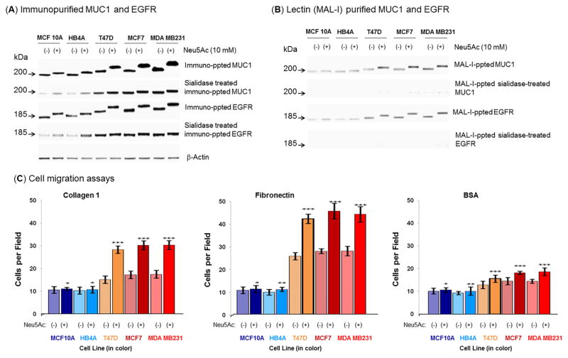
Figure 6. Examination of sialylation of MUC1 and EGFR on normal and malignant cells after sialic acid treatment under nutrient deprivation and cell migration.
(A) Equal amount (100 μg) of each cell extract was subjected to immuno-precipitation with anti-MUC1 and anti-EGFR antibodies and the precipitated proteins were subjected to Western blot and immuno-detection with the respective antibody. In parallel, equal amount of each crude protein extract was desialylated and similar precipitation was carried out. (B) Equal amount of immuno-precipitated proteins (MUC1 and EGFR) as described above was subjected to MAL-I precipitation and detected on W. blot as described above. (C) Cell migration. Migration assays were performed on confluent monolayers treated or not treated with 10 mM Neu5Ac under nutrient deprived condition. Data are shown as the number of cells that migrated into a 300×300 micron area along the center of the wound in 24 hours. The data are representative of five independent experiments with S.D. indicated by error bars. P values are determined by two-tailed student t-test and <0.05, <0.01 and <0.001 are indicated by one, two and three asterisks, respectively compared to the corresponding untreated cells.

