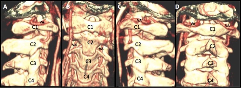
Figure 3. Tri-dimensional and Multiplanar Reconstructions of the Occiputo-Cervical Region Including VA and C1-C4 Vertebral Bodies.
Tri-dimensional and multiplanar reconstructions of the occiputo-cervical region, including VA and C1-C4 vertebral bodies in lateral, AP, and posterior projections (Panels A-D). By importing the computed tomography angiography images into any system with the tri-dimensional reconstruction capability, we will be able to visualize the occiputo-cervical bony structures with the VA in place. This gives us a 3D image of the position of the vertebral artery before starting screw fixation.

