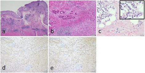
Fig. 7.

Histological findings. a Hematoxylin-eosin staining (×20), cancer cells spread horizontally at submucosal layer. b Hematoxylin-eosin staining (×100), marked infiltration of the plasma cells and lymphocytes was shown at tumor stroma. c Hematoxylin-eosin staining (×400), infiltration of plasma cells and fibrosis were shown in the retroperitoneal tissue. d IgG-immunostaining (×100) and e IgG4-immunostaining (×100), more than half of the infiltrating plasma cells were positive for IgG4
