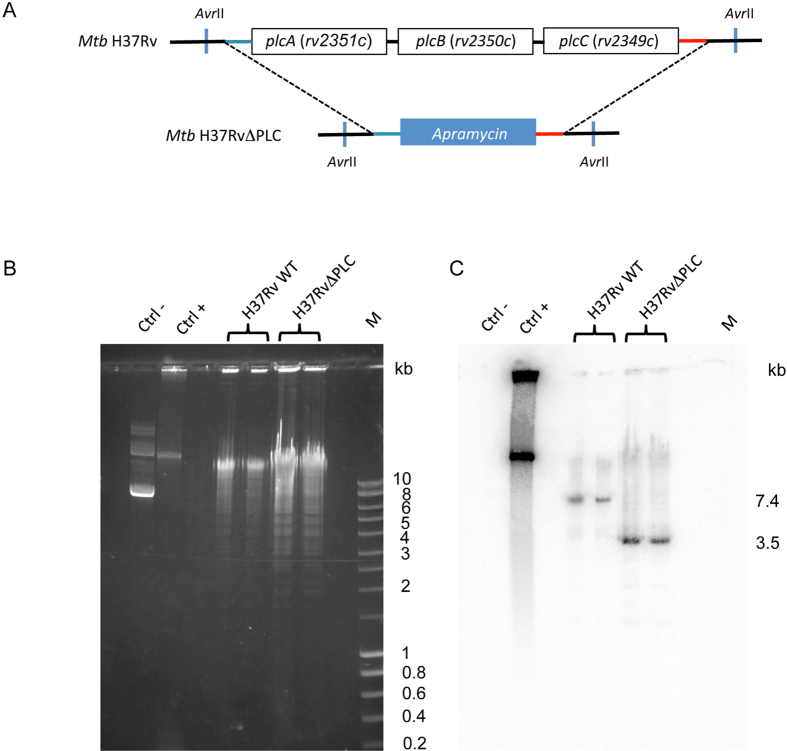
Figure 1. Construction of M. tuberculosis H37RvplcABC KO (H37RvΔPLC).
(A) Schematic representation of genomic organization of plc genes in M. tuberculosis H37Rv wild type and H37RvΔPLC strains ; (B) AvrII restriction fragment profiles of M. tuberculosis WT and KO strains separated by agarose gel electrophoresis; (C) Pattern obtained from genomic DNAs digested with AvrII and hybridized with a probe specific for the plcC downstream region; Lanes: 1 (second lane from left), negative control (pYUB412 vector); 2, positive control pYUB412::plcABC; 3 and 4, M. tuberculosis H37Rv WT, 5 and 6, M. tuberculosis H37RvΔplcABC; 7, M, Smart Ladder (Eurobio).

