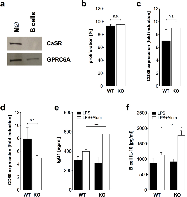
Figure 6. In vitro activation of B cells by LPS/Alum induces increased antibody production in GPRC6A−/− B cells.
(a) Immunoblot analysis demonstrating the expression of CaSR and GPRC6A in peritoneal macrophages (M∅) and splenic B cells isolated from C57/BL6 mice. Shown is one representative experiment out of three. (b–d) Splenic B cells from 4 wildtype (WT, black) and 3 GPRC6A−/− mice (KO, white) were in vitro activated with LPS/Alum and proliferation on day 7 (b), CD86 expression on day 7 (c) and CD69 expression on day 1 (d) was analyzed by flow cytometry. Statistical analysis was performed using t-test. Bars represent mean ± SEM. (e) Splenic B cells from 3 wildtype (WT, black, n = 9) and 3 GPRC6A−/− mice (KO, white, n = 7) were in vitro activated with LPS or LPS/Alum and IgG1 concentration in the supernatant was analyzed by ELISA on day 7. Statistical analysis was performed using t-test. Bars represent mean ± SEM. (f) Splenic B cells from 2 wildtype (WT, black, n = 6) and 2 GPRC6A−/− mice (KO, white, n = 6) were in vitro activated with LPS or LPS/Alum and IL-10 concentration in the supernatant was analyzed by ELISA on day 7. Statistical analysis was performed using t-test. Bars represent mean ± SEM. (**P < 0.01, ***P < 0.001).

