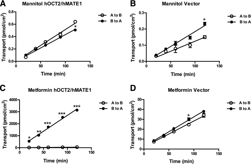
Fig. 6.
Transcellular flux of mannitol and metformin in hOCT2/hMATE1 double-transfected and control MDCK cells. Cells were incubated in KRH buffer containing 0.05 μM mannitol (A and B) and 5.5 μM metformin (C and D) in either the apical (open circle) or basal (closed circle) chamber. An aliquot of buffer was taken periodically from the receiving chamber (100 μl from the basal chamber and 50 μl from the apical chamber) and radioactivity was subsequently measured. The pH of the apical and basal chambers was 6.0 and 7.4, respectively. Data were fitted with linear regression. Each data point represents the mean ± S.D. Transport in the basal-to-apical direction was compared with that in the apical-to-basal direction (*P < 0.05; **P < 0.01; ***P < 0.001). A to B, apical to basal; B to A, basal to apical.

