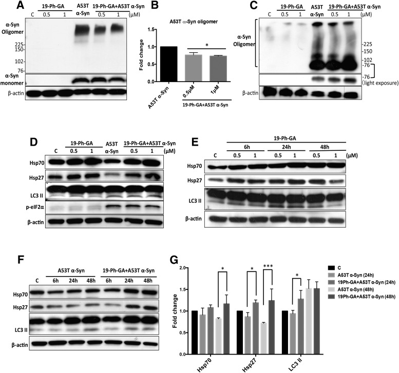
Fig. 6.
19-Ph-GA significantly reduced A53T α-Syn oligomer formation through induction of heat shock protein responses and autophagy. (A and B) α-Syn oligomers (denatured) were significantly reduced after 48-hour post-treatment of 5Y cells with 19-Ph-GA. (B) Fold changes of α-Syn oligomers in (A) after the drug treatment are normalized to respective controls and estimated by densitometry. (C) Exposure of 5Y cells to 19-Ph-GA resulted in decreased levels of native high molecular weight α-Syn oligomers as well as an increase in the smaller (76 kDa) oligomer species. (D) After 48-hour post-treatment with 19-Ph-GA, Hsp70 and Hsp27 protein levels were elevated 2- to 3-fold, but biomarkers of autophagy and ER stress were not significantly perturbed. (E) Time-course study showed that 19-Ph-GA was able to upregulate both HSPs and LC3 II expression in 5Y cells. Note that exposure to 19-Ph-GA for 24 hours induced the maximum increase in LC3 II, suggesting 19-Ph-GA stimulated a temporal autophagic flux in 5Y cells. (F and G) Significant induction of HSPs and LC3 II could be detected in A53T α-Syn–overexpressing cells after post-treatment with 19-Ph-GA (0.5 μM) for 24 hours, suggesting that HSP response and autophagic flux were stimulated by 19-Ph-GA at relatively early time points. β-Actin was included as a loading control. A representative blot from three separate experiments is shown for each figure. (G) Fold changes of Hsp70, Hsp27, and LC3 II in (F) after the drug treatment at the indicated times are normalized to respective controls and estimated by densitometry. Values in (B) and (G) are presented as the mean ± S.D. (n = 3); *P < 0.05 considered significant compared with the A53T α-Syn group by one-way analysis of variance using Tukey’s multiple comparison test. p-eIF2α, phosphorylated eIF2α.

