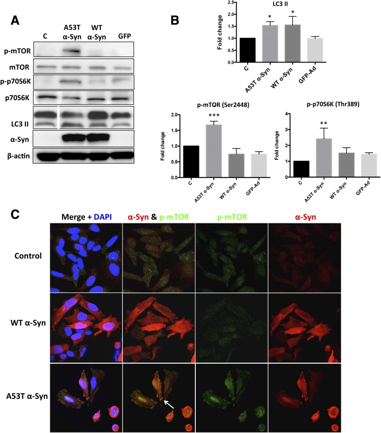
Fig. 7.
mTOR/p70S6K signaling was activated by overexpression of A53T but not WT α-Syn. (A) Overexpression of A53T, but not WT for 72h, α-Syn significantly increased the levels of phosphorylated mTOR and p70S6K in 5Y cells. An increased amount of LC3 II was observed in both A53T and WT α-Syn–overexpressing cells, suggesting that autophagy was not directly mediated via the mTOR/p70S6K signaling. The white dividing line in the LC3 II blot indicates omission of three lanes on the gel; all lanes shown were from the same gel. (B) Fold changes of LC3 II, p-mTOR, and p-p70S6K levels in (A) are normalized to respective controls and estimated by densitometry. These values are presented as the mean ± S.D. (n = 3); *P < 0.05 is considered significant relative to the control group using Dunnett’s multiple comparison test. (C) Increased expression of p-mTOR was observed in A53T, but not WT, α-syn–overexpressing cells. After transduction with adenovirus expressing A53T α-Syn or WT α-Syn at MOI 400 plaque-forming units/cell for 72 hours, 5Y cells were fixed and immunostained for α-Syn (red), p-mTOR (green), and nuclei (DAPI, blue) and then examined by confocal microscopy. Note that the colocalization of p-mTOR and α-Syn (white arrow), suggesting an interaction between p-mTOR and A53T α-Syn, may exist. Ad, adenovirus; GFP, green fluorescent protein; DAPI, 4′,6-diamidino-2-phenylindole; MOI, multiplicity of infection.

