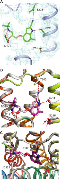
Fig. 6.
Structure of 7-methylcyanopindolol–bound β1AR. (A) Omit map for the ligand-binding site. A 2Fo-Fc map was generated where the ligand and side chains of Ser211 and Ser215 were omitted from the model. The contour level is 1.0 sigma, and the figure was produced using CCP4MG. (B and C) Superposition of the structures of β1AR bound to 7-methylcyanopindol (receptor in rainbow coloration and the ligand in pink) and cyanopindol (receptor and ligand are both gray). The views are either within the membrane plane (A and B) or from the extracellular surface (C), with portions of H3 removed for clarity. The red arrows highlight the different positions of Ser215 in the two β1AR structures. The Ballesteros-Weinstein numbers for the residues depicted are as follows: D121, 3.32; S211, 5.42; S215, 5.46; and N329, 7.39.

