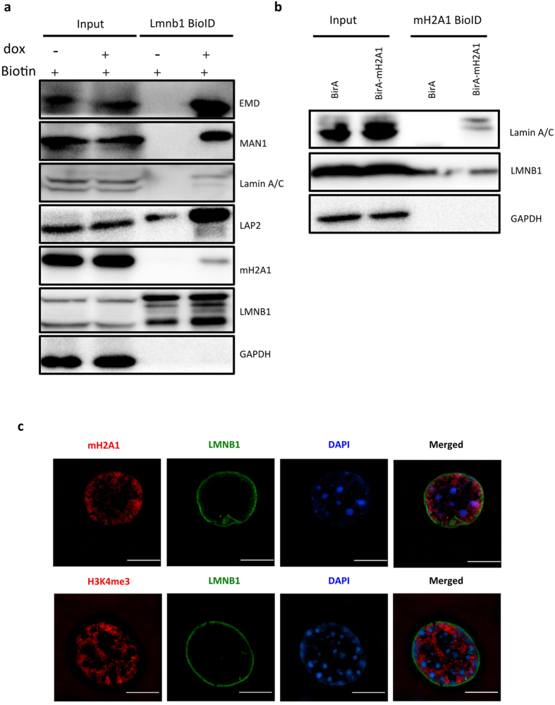
Figure 2. LMNB1 associates with histone variants macroH2A1.
(a) Validation of proteomic data with western blotting. Whole cell extract (Input) and captured biotinylated proteins (BioID) were blotted with the indicated antibodies, in presence of biotin. Starting materials were equal for the samples with (+) and without (−) dox. GAPDH was used as a negative control. (b) Test of macroH2A1-LMNB1 association by BioID using macroH2A1 as the bait. Whole cell extract (Input) and macroH2A1 BioID products in Hela cells were blotted with the indicated antibodies. GAPDH was used as a negative control. (c) Sub-cellular localization of macroH2A1 or H3K4me3 (red) and LMNB1 (green) were shown by immunofluorescence and super-resolution microscope (SIM) in AML12. DNA was labeled with DAPI (blue). Scale = 10 μm.

