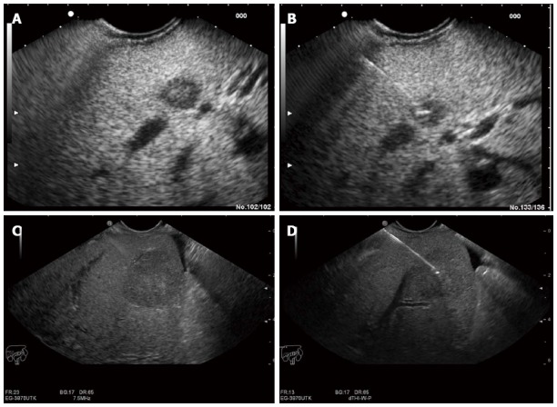
Figure 5.

Endoscopic ultrasound image of lesion in the liver. A: Endoscopic ultrasound (EUS) image of an 8 mm metastatic lesion in the liver; B: Endoscopic ultrasound (guided biopsy from the same lesion; C: EUS image of a 25 mm lesion in the liver; D: EUS guided aspiration biopsy from the same lesion.
