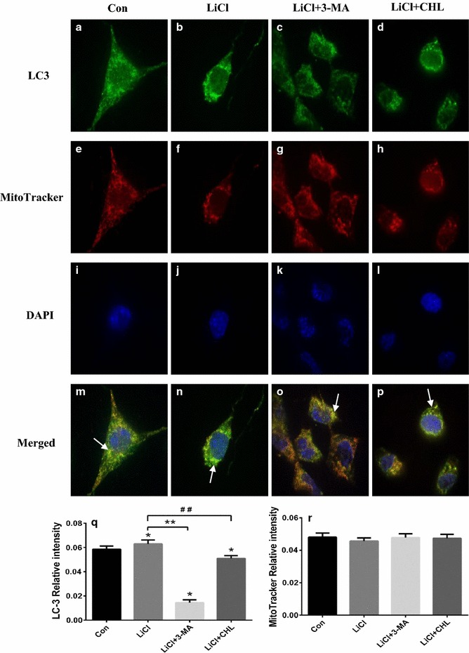
Fig. 5.

Lithium promoted degradation of mitochondria via autophagy. Cells exposed to lithium with or without autophagy inhibitors were incubated with anti-LC3 antibody (1:100) and MitoTracker Red to visualize the co-localization (yellow immunofluorescence) of LC3 (a–d) and mitochondria (e–h) in SH-SY5Y cells at ×400 magnifications under a confocal fluorescence microscope, and the nuclei were stained by DAPI (i–l). The co-localization of autophagosomes and mitochondria was marked using white arrows (m–p). The relative intensity of LC3 (q) and mitochondria were expressed (r). *P < 0.05 as compared with Con group; **P < 0.05 as compared with LiCl + Rot group; ## P < 0.05 as compared with LiCl + Rot group (n = 6 in each group)
