
Figure 3.
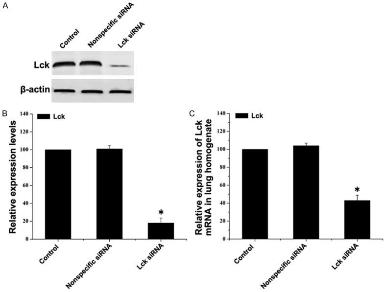
In vivo transfection of Lck siRNA reduces Lck expression in mouse lung tissue. Mice were sensitized with OVA and transfected via tail vein injection with Lck siRNA or nonspecific siRNA and then challenged with OVA. Control mice were sensitized and challenged with PBS and not transfected. Mice were sacrificed and lung tissue was removed following the final OVA challenge. A. Expression levels of Lck in lung tissue were detected by western blot. β-actin served as the loading control. B. Quantitative analysis of the changes of Lck. Data represent mean ± SEM (n=3, *P < 0.05 vs control group). C. Total RNA was extracted from lung and expressions of Lck mRNA levels were detected by quantitative real-time PCR. The amount of Lck mRNA relative to β-actin is shown. Data represent mean ± SEM (n=3, *P < 0.05 vs control group).